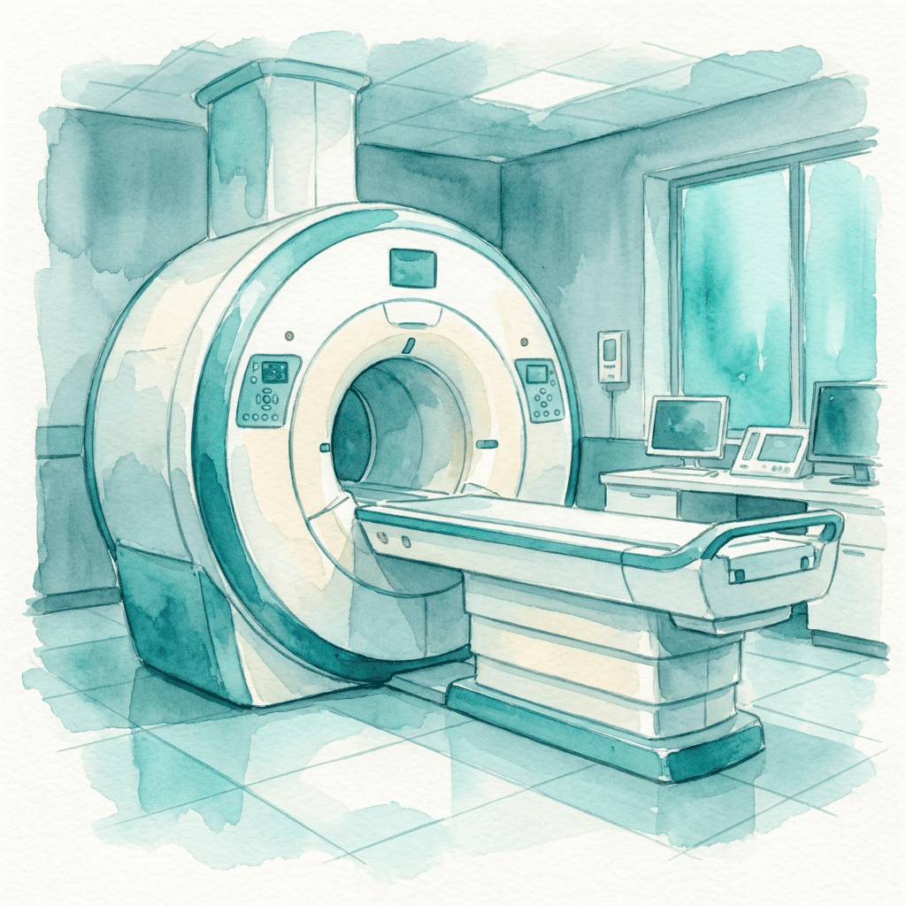

Beyond the Standard MRI
You finally got a doctor to order an MRI. A few days later the report came back: no acute findings. Within normal limits.
You read it twice. How can your head constantly ache and throb, yet nothing is wrong?
If you’ve been holding a scan that was called normal while your body keeps telling you something is wrong, you’re not imagining the gap between those two things.
What I Learned About Imaging
I had been scanned before, and every MRI report came back normal. Then I had four different scans: Digital Motion X-ray (DMX), Upright MRI, cone beam CT, and prone lumbar MRI. Each one showed something the standard MRI couldn’t.
Standard Supine MRI
This is the scan most people get. The patient lies flat and the body is supported. The spine is in its most stable position. The machine takes detailed pictures of soft tissue, bone, and fluid, and it catches structural problems that exist regardless of position, such as tumors, large herniations, fractures, and lesions.
What it can’t show is anything that depends on gravity, motion, or upright posture to become visible.
It doesn’t see what happens when the body works.
Digital Motion X-ray (DMX)
A DMX is usually performed while standing, which captures how the joints function under the weight of gravity. It records movement in real time, allowing instability that appears during motion to become visible.
Mine showed overhang at multiple levels, making visible what my standard supine imaging had missed.
Upright MRI
An upright MRI is performed with the patient seated or standing.
When a person lies down, the cerebellum can sit higher in the skull than it does when they’re upright. A Chiari malformation that’s visible standing can sometimes appear reduced or disappear lying down.
Mine showed Chiari and confirmed the instability the DMX had already captured.
Cone Beam CT
A cone beam CT is a high-resolution scan of the bony anatomy. Where MRI shows soft tissue, fluid, and structural relationships, cone beam CT shows bone in fine detail.
A cone beam CT can also capture alignment in different positions, including when the head bends forward, tilts back, or turns side to side.
Prone Lumbar MRI
A standard lumbar MRI is performed face-up. A prone lumbar MRI flips the patient face-down, changing how tension appears along the spinal cord.
Mine showed tethered cord that none of my face-up lumbar scans had caught.
If You’re Already Asking About These Scans
You may be reading this because you’ve heard about upright MRI or DMX through a forum, social media group, or another patient.
Before you start searching for these scans, a few realities are worth knowing:
• Most providers are not aware that these scans exist
• Not every imaging center has the equipment
• Many radiologists aren’t trained to interpret them
If you have been told for years that your imaging is fine while your body tells you something else, the explanation you have been given may not be the only one. The right scan is only part of the puzzle. The other part is finding the clinician who knows how to interpret it.
Want to understand the structural cause?
Craniocervical Instability (CCI): A Patient’s Perspective.
Every stripe tells part of the story.
The full story begins in You Might Be a Zebra: Rare Unveiled.


Please note: This information is provided for general reference only and is not based on medical advice, diagnosis, or treatment. Always consult a qualified healthcare professional for medical concerns.